Microscope Parts and Functions: A Comprehensive Guide
Exploring microscopic worlds demands understanding key components.
From eyepieces to objective lenses, and illumination systems,
a microscope’s function relies on precise parts.
PDF guides detail SEM, TEM, and field-ion microscopy,
highlighting their unique roles in scientific observation and analysis.
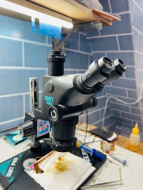
Microscopy, the technical field of using microscopes to view objects or details that are too small to be seen by the naked eye, has revolutionized numerous scientific disciplines. Understanding the parts and functions of a microscope is crucial for effective observation and analysis. Traditional light microscopes, detailed in readily available PDF guides, consist of essential components like the eyepiece (ocular lens), objective lenses, and a robust microscope stand.
Beyond basic light microscopy, advanced techniques such as phase contrast microscopy and dark field microscopy offer enhanced visualization. For even greater magnification and resolution, electron microscopy – including Scanning Electron Microscopy (SEM) and Transmission Electron Microscopy (TEM) – becomes essential, particularly when examining structures below 0.2 micrometers. Specialized methods like Emission Microscopy (EMMIE) further expand observational capabilities. These techniques, and their corresponding components, are comprehensively documented in specialized literature and downloadable PDF resources.
Types of Microscopes
Microscopes extend our vision into realms unseen by the naked eye, categorized by their underlying principles. Light microscopes, the most common type, utilize visible light and lenses – their parts and functions are thoroughly explained in numerous PDF guides. However, when investigating structures beyond the resolution limits of light, electron microscopes become indispensable.
Scanning Electron Microscopy (SEM) provides detailed images of surfaces, while Transmission Electron Microscopy (TEM) reveals internal structures. More specialized techniques include field-ion microscopy, focusing on atomic-level observation, and Emission Microscopy (EMMIE), used for semiconductor analysis. Each type boasts unique parts – electron guns, vacuum systems, and specialized detectors – and distinct functions. Comprehensive PDF resources detail the operational principles, component breakdowns, and applications of each microscopic modality, aiding in informed selection for specific research needs.
Basic Components of a Light Microscope
A standard light microscope, detailed in accessible PDF guides, comprises several key components working in harmony. The microscope stand provides structural support, housing the optical components – eyepiece (ocular lens) and objective lenses – responsible for magnification. Precise positioning is achieved via stage and stage controls, allowing for systematic sample observation.
Crucially, focusing knobs (coarse and fine) enable sharp image acquisition. The illumination system, including the light source and condenser with iris diaphragm, controls light intensity and contrast. Understanding the parts and their respective functions is paramount for effective microscopy. Numerous PDF resources offer detailed diagrams and explanations, covering assembly, operation, and maintenance, ensuring optimal performance and extending the microscope’s lifespan.
Optical Components
The optical components are central to a microscope’s image-forming process, thoroughly explained in detailed PDF manuals; The eyepiece (ocular lens) magnifies the image produced by the objective lenses, typically offering 10x magnification. Objective lenses, ranging from 4x to 100x, provide varying levels of magnification and resolution, crucial for observing different sample details.
These lenses are designed to correct for optical aberrations, ensuring a clear and accurate image. PDF guides emphasize proper lens care and selection based on the specimen. Understanding the numerical aperture (NA) of each objective is vital for maximizing resolution. Together, these parts work to gather light and create a magnified visual representation, forming the foundation of microscopic observation, as detailed in comprehensive function guides.
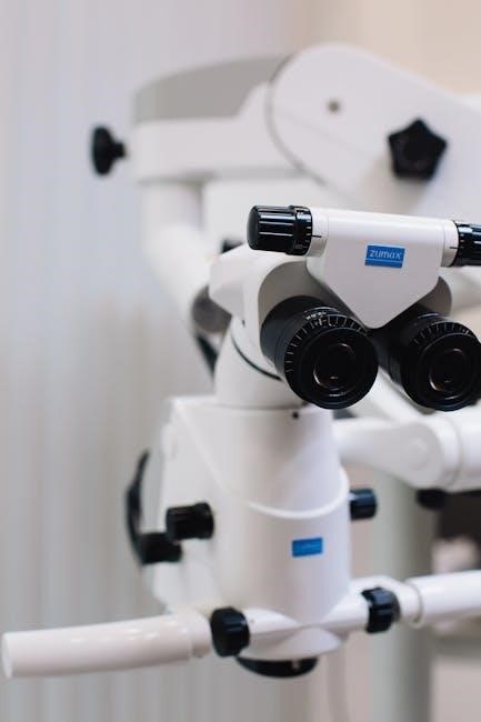
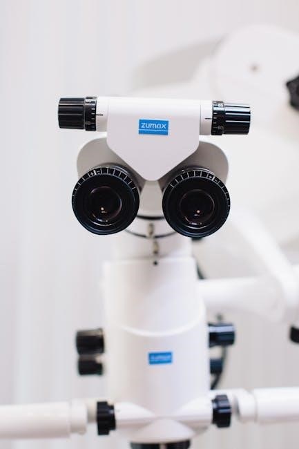
The Eyepiece (Ocular Lens)
The eyepiece, also known as the ocular lens, is the lens through which the observer views the specimen, detailed in numerous PDF resources. Typically magnifying the image by 10x, it further enlarges the image created by the objective lens. PDF guides often explain different eyepiece types, including those with adjustable diopters to compensate for individual vision differences.

Its primary function is to provide a comfortable viewing experience and enhance magnification. Some eyepieces feature reticles – scales used for measuring specimen dimensions. Proper alignment of the eyepiece is crucial for optimal image clarity, as outlined in maintenance PDFs. Understanding its role is fundamental to mastering microscopy, and detailed diagrams are readily available for download as parts references.
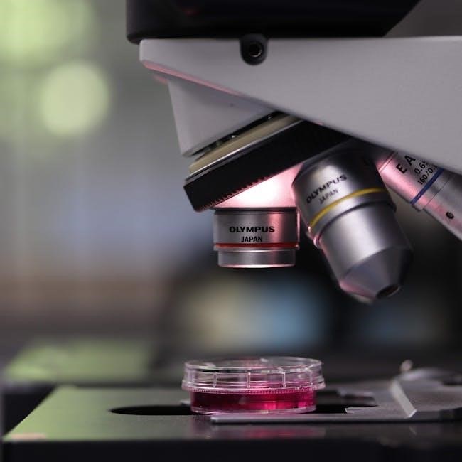
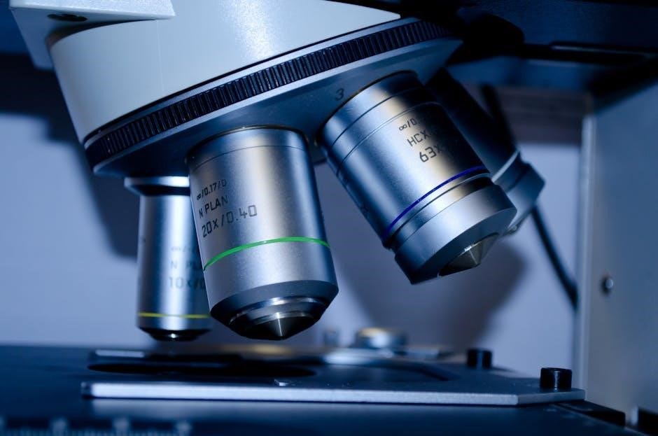
Objective Lenses
Objective lenses are the most important part of any microscope, responsible for the primary magnification and resolution of the specimen – details extensively covered in microscopy PDF guides. These lenses are mounted on a revolving nosepiece, allowing for quick changes in magnification power, typically ranging from 4x to 100x. PDF resources often categorize them by their numerical aperture (NA), a measure of light-gathering ability.
Their function is to collect light from the specimen and create a magnified image. Higher magnification objectives require greater illumination and are often used with immersion oil to improve resolution, as explained in advanced technique PDFs. Understanding the parts – the lens elements and their arrangement – is key to proper use and maintenance, readily available in downloadable manuals.
Mechanical Components

Mechanical components provide the structural support and control for a microscope, crucial for stable observation – details often found in comprehensive microscope PDF guides. The microscope stand, or base, offers stability, while the stage securely holds the specimen. Stage controls allow precise movement in both horizontal (X and Y) directions, enabling systematic scanning of the sample, as illustrated in many instructional PDFs.
Focusing knobs – coarse and fine – are essential for achieving a clear image. Coarse adjustment provides large-scale focusing, while fine adjustment allows for precise sharpening. Detailed diagrams of these parts and their function are readily available in downloadable manuals and PDF resources, ensuring proper operation and maintenance for optimal performance.
The Microscope Stand
The microscope stand, often referred to as the base, forms the foundational support for all other components. Detailed schematics within microscope PDF guides emphasize its robust construction, typically made of metal for stability. It houses the illumination system and supports the stage and body tube. A stable stand minimizes vibrations, crucial for high-magnification observation.
Many PDF resources highlight the stand’s ergonomic design, ensuring comfortable and prolonged use. Some stands include built-in features like adjustable feet for leveling on uneven surfaces. Understanding the stand’s function – providing a secure and stable platform – is fundamental to proper microscope operation, as explained in numerous instructional PDFs detailing microscope parts.
Stage and Stage Controls
The stage is the flat platform where specimens are placed for observation. Microscope PDF guides detail various stage types, including mechanical stages offering precise X-Y movement via stage controls. These controls, often knobs, allow for smooth and accurate positioning of the slide, essential for scanning and selecting areas of interest. Some advanced stages, illustrated in detailed PDF diagrams, incorporate vernier scales for precise measurements.
Stage clips secure the slide, while focusing on PDF resources emphasizes the importance of clean stages for optimal imaging. Understanding the function of each control – moving the slide horizontally and vertically – is crucial. PDF manuals often include troubleshooting tips for sticky or unresponsive stage controls, ensuring proper operation of this vital microscope part.
Focusing Knobs (Coarse and Fine)
Focusing knobs are essential for achieving a clear image. Microscope PDF guides clearly illustrate the difference between coarse and fine focus. The coarse adjustment knob allows for large-scale adjustments to the stage height, quickly bringing the specimen into approximate focus – PDF resources caution against overuse with high-power objectives. The fine focus knob, detailed in many PDF manuals, provides precise adjustments for sharp, detailed imaging.
Understanding their function is critical; PDF diagrams show how they manipulate the objective lens position. Proper use, as explained in PDF tutorials, prevents damage to the objective and slide. Troubleshooting sections in PDF documents address issues like stiff knobs or inability to achieve focus, highlighting the importance of regular maintenance of these key microscope parts;
Illumination System
The illumination system is vital for visualizing specimens, and detailed PDF guides explain its components. This system comprises the light source and the condenser with its iris diaphragm. PDF resources emphasize the light source’s role in providing necessary illumination, ranging from halogen lamps to LEDs. The condenser, as illustrated in microscope parts PDFs, focuses light onto the specimen, enhancing resolution and contrast.
Adjusting the iris diaphragm, covered in many PDF tutorials, controls the amount of light reaching the sample, optimizing image clarity. Proper illumination, detailed in PDF maintenance guides, is crucial for various microscopy techniques. PDF troubleshooting sections address issues like uneven illumination or flickering lights, ensuring optimal performance of these essential microscope parts and their function.
The Light Source
The light source, a fundamental component detailed in microscope parts PDF guides, provides the illumination necessary to view specimens. Historically, PDF resources show, halogen lamps were common, but modern microscopes increasingly utilize LEDs for their efficiency and longevity. These PDF guides explain how the light source’s intensity is adjustable, impacting image brightness and contrast. Understanding the function of the light source is crucial for optimal microscopy.
PDF maintenance manuals emphasize proper handling and replacement procedures for the light source. Troubleshooting sections within these PDFs address common issues like bulb burnout or inconsistent illumination. Detailed diagrams in parts lists PDFs illustrate the light source’s placement within the overall illumination system, highlighting its integral function in producing a clear, visible image.
Condenser and Iris Diaphragm
Microscope parts PDF guides extensively cover the condenser and iris diaphragm, vital for controlling light quality. The condenser, as detailed in these PDFs, focuses light onto the specimen, enhancing resolution and contrast. Adjustment of the condenser height is crucial, a point emphasized in many instructional PDFs. The iris diaphragm, also explained in parts diagrams within PDFs, regulates the amount of light reaching the specimen.
These PDF resources illustrate how manipulating the iris diaphragm affects image clarity and depth of field. Proper function of both components is essential for optimal viewing. Troubleshooting sections in maintenance PDFs address issues like a hazy condenser or a stuck iris diaphragm. Understanding their combined function, as shown in exploded view PDFs, is key to effective microscopy.
Advanced Microscopy Techniques
PDF guides detailing microscope parts and function often extend to advanced techniques. Phase contrast microscopy, explained in specialized PDFs, enhances contrast in transparent specimens without staining, crucial for live cell observation. Dark field microscopy, also covered in these resources, illuminates specimens with oblique light, revealing details invisible under standard conditions. These techniques rely on modifications to the standard microscope setup, detailed in component parts lists within PDFs.
Further PDF documentation explores techniques like emission microscopy (EMMIE), utilizing photon detection for semiconductor analysis. Understanding the function of each technique requires knowledge of specific optical components, as illustrated in schematic PDFs. Maintenance PDFs also address calibration procedures for these advanced systems, ensuring accurate results. These advanced methods expand the capabilities of microscopy beyond basic observation.
Phase Contrast Microscopy
Phase contrast microscopy, thoroughly explained in dedicated PDF guides, leverages differences in refractive index within transparent samples. These PDFs detail the crucial parts – a special annular diaphragm in the condenser and a phase plate within the objective lens. The annular diaphragm shapes the illuminating light, while the phase plate alters the phase of light passing through the specimen. This creates visible contrast from otherwise transparent structures.
PDF documentation emphasizes the function of these components: the phase plate shifts the phase of diffracted light, enhancing visibility. Detailed diagrams in these guides illustrate light paths and component placement. Proper alignment, as outlined in maintenance PDFs, is vital for optimal image quality. Understanding these parts and their function is key to successful phase contrast imaging, allowing visualization of live cells and unstained specimens.
Dark Field Microscopy
Dark field microscopy, comprehensively covered in specialized PDF resources, creates a distinctive image by illuminating the sample with light that does not directly enter the objective lens. These PDF guides detail the essential parts: a powerful light source, a specialized dark field condenser, and a standard objective lens. The condenser blocks direct light, allowing only scattered light from the specimen to reach the observer.
The function of this technique, explained in detail within these PDFs, is to reveal unstained, transparent specimens. Scattered light appears bright against a dark background, highlighting details invisible under brightfield. PDFs often include troubleshooting tips for optimal setup and image clarity. Understanding the interplay of these parts and their function is crucial for observing living microorganisms, blood cells, and other delicate samples without staining.
Electron Microscopy
Electron microscopy, extensively detailed in dedicated PDF guides, utilizes beams of electrons instead of light to create highly magnified images. These PDF resources outline the core parts: an electron source (like a filament), electromagnetic lenses to focus the beam, and a viewing screen or detector. Understanding each part’s function is vital for successful operation.
The primary function of electron microscopy, as explained in these PDFs, is to achieve resolutions far beyond the capabilities of light microscopy. Two main types are covered: Scanning Electron Microscopy (SEM), which images surface topography, and Transmission Electron Microscopy (TEM), revealing internal structures. PDFs emphasize sample preparation techniques and image interpretation. These guides also detail the vacuum systems essential for electron beam travel, ensuring optimal image quality and resolution.
Scanning Electron Microscopy (SEM)
Scanning Electron Microscopy (SEM), thoroughly explained in specialized PDF guides, creates images by scanning a focused electron beam across a sample’s surface. These PDFs detail the key parts: an electron gun, condenser lenses to focus the beam, scanning coils to raster the beam, and detectors to capture emitted signals. Understanding each part’s function is crucial for optimal imaging.
The primary function of SEM, as outlined in these PDF resources, is to produce high-resolution images of surface topography and composition. Samples often require conductive coating. PDFs emphasize signal detection methods – secondary electrons for topographical contrast and backscattered electrons for compositional information. These guides also cover vacuum systems vital for electron beam travel and image quality, alongside detailed explanations of image formation and interpretation.
Transmission Electron Microscopy (TEM)
Transmission Electron Microscopy (TEM), comprehensively detailed in available PDF resources, forms images by transmitting a beam of electrons through an ultra-thin specimen. These PDF guides meticulously explain the core parts: an electron gun, condenser lens system, objective lens, and image projection system. Understanding each part’s function is paramount for successful operation.
The primary function of TEM, as elucidated in these PDFs, is to reveal internal structures at the nanometer and even atomic scales. Sample preparation—requiring extremely thin sections—is a critical step, detailed in many PDFs. These resources explain how electrons interact with the sample, creating contrast based on density and composition. PDFs also cover the importance of vacuum systems and image interpretation techniques, offering a complete understanding of this powerful microscopy method.

Field-Ion Microscopy
Field-Ion Microscopy (FIM), thoroughly explained in specialized PDF guides, is a unique technique visualizing surfaces at atomic resolution. These PDF resources detail the core parts: a sharply pointed metallic tip, a high-voltage power supply, and a fluorescent screen or detector. Understanding each part’s function is crucial for operation.
The primary function of FIM, as outlined in these PDFs, involves applying a strong electric field to the tip, ionizing surface atoms. These ions are then projected onto a screen, forming an image. PDFs emphasize the importance of ultra-high vacuum conditions and precise tip preparation. They also explain how image contrast relates to the local work function of the surface atoms. Detailed PDFs cover the analysis of these images, revealing surface defects and atomic arrangements, offering insights unavailable with other techniques.
Specialized Microscopy Techniques
Specialized microscopy techniques, comprehensively documented in detailed PDF guides, extend beyond conventional methods. These PDF resources explore techniques like Emission Microscopy (EMMIE), focusing on semiconductor device analysis. Understanding the parts and their function is vital for researchers.
PDFs explain that EMMIE utilizes a high-gain camera to capture photons emitted from defects within semiconductors under voltage or current. These guides detail the importance of precise voltage control and detector sensitivity. Other PDFs cover techniques for analyzing material properties, often requiring specialized sample preparation and image processing. They emphasize the role of each component – from the light source to the detector – in achieving optimal results. Accessing these PDFs provides a deeper understanding of advanced imaging capabilities.
Emission Microscopy (EMMIE)
Emission Microscopy (EMMIE), thoroughly explained in specialized PDF guides, is a powerful technique for analyzing semiconductor devices. These PDFs detail how EMMIE captures faint light emitted by defects or failures within the semiconductor material. Understanding the parts and their function is crucial for effective analysis.
PDF documentation highlights the use of high-gain cameras or detectors to capture these weak photon emissions. Applying a voltage or current to the sample accelerates charge carriers, generating light revealing localized issues. The PDFs emphasize the importance of precise control over applied voltage and the detector’s sensitivity. They also cover image processing techniques to enhance visibility. Accessing these PDF resources provides a detailed understanding of EMMIE’s capabilities and limitations, aiding in semiconductor failure analysis.
Microscope Maintenance and Care
Maintaining optimal performance, as detailed in comprehensive PDF guides, is vital for any microscope. These PDFs emphasize regular cleaning of all parts – lenses, stage, and illumination system – using appropriate lens paper and cleaning solutions. Understanding each component’s function aids in targeted care.
PDF resources outline proper storage procedures, protecting the microscope from dust, humidity, and extreme temperatures. They also detail lubrication points for mechanical parts, ensuring smooth operation. Regular checks of the illumination system, including the light source and condenser, are crucial. Following the guidelines in these PDFs extends the microscope’s lifespan and ensures accurate, reliable results. Preventative maintenance, as described, minimizes costly repairs and downtime, maximizing the instrument’s utility.
Troubleshooting Common Microscope Issues
Comprehensive PDF guides are invaluable when addressing microscope malfunctions. These resources detail solutions for common problems, linking issues to specific parts and their functions. Blurred images often stem from dirty objective lenses or improper focusing, explained clearly in PDFs.
PDFs also address illumination problems – dim light, uneven brightness – often related to the light source or condenser. Mechanical issues, like stage movement difficulties, are linked to lubrication or alignment, with step-by-step fixes. Understanding the function of each component, as outlined in the PDFs, aids diagnosis. These guides emphasize safety precautions during troubleshooting. Regularly consulting these PDF resources minimizes downtime and ensures accurate observations, extending the microscope’s operational life.
Digital Microscopy and Image Capture
Modern microscopy increasingly integrates digital imaging, detailed in specialized PDF guides. These resources explain connecting cameras – SEM, TEM compatible – to microscopes, outlining the function of each interface. PDFs cover software for image acquisition, processing, and analysis, crucial for documenting observations.
Understanding the parts involved – adapters, cameras, computers – is vital, and PDFs provide schematics and troubleshooting tips. They explain optimizing settings for different objectives and illumination techniques. Digital microscopy enhances image clarity and allows for measurements and annotations. PDF guides also address file formats, resolution, and storage. Mastering these digital techniques, as detailed in the PDFs, expands research capabilities and facilitates data sharing.
Applications of Microscopy in Various Fields
Microscopy’s versatility, detailed in comprehensive PDF guides, extends across numerous disciplines. In materials science, techniques like SEM analyze surface morphology, while TEM reveals internal structures – information readily available in downloadable PDFs. Biological research utilizes light microscopy to study cells, and specialized PDFs explain techniques like phase contrast.
Medical diagnostics rely on microscopy for identifying pathogens and analyzing tissue samples; relevant PDFs cover staining methods and image interpretation. Forensic science employs microscopy for trace evidence analysis, with PDF resources detailing comparative microscopy. Understanding the parts and function of each microscope type, as outlined in these PDFs, is crucial for accurate results. These guides demonstrate microscopy’s broad impact, showcasing its essential role in scientific advancement.
Resources for Further Learning (PDF Guides)
Numerous PDF guides offer in-depth exploration of microscope parts and their functions. These resources detail everything from basic light microscopy to advanced techniques like SEM and TEM, providing schematics and operational instructions. Many university websites host free PDFs covering optical component alignment and troubleshooting. Commercial microscope manufacturers also provide downloadable PDF manuals and application notes.
Specialized PDFs focus on specific microscopy methods, such as phase contrast or dark field illumination, explaining the principles and practical implementation. Guides detailing field-ion microscopy and emission microscopy (EMMIE) are also available. These PDFs often include safety precautions and maintenance schedules, ensuring responsible microscope use. Accessing these resources enhances understanding and maximizes the potential of microscopic observation.
Safety Precautions When Using a Microscope
Prioritize safety when operating a microscope. Always ensure the light source is adjusted to a comfortable intensity to prevent eye strain. Handle slides and coverslips with care to avoid breakage and potential cuts – dispose of broken glass properly. When using oil immersion objective lenses, only use immersion oil specifically designed for microscopy, and clean lenses thoroughly afterward.

Be mindful of electrical safety; ensure the microscope is properly grounded and avoid contact with water near electrical components. When exploring advanced techniques like SEM or TEM (detailed in accompanying PDF guides), adhere to specific radiation safety protocols. Never disassemble microscope parts without proper training. Regular maintenance, as outlined in manufacturer PDFs, contributes to safe operation.